OUR GUIDE TO CERVICAL CANCER:
CERVICAL SCREENING AWARENESS WEEK 15-21 JUNE 2020
Cervical cancer is the most common form of cancer in women under 35 in the UK with
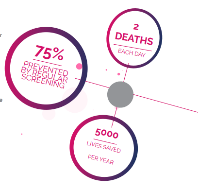
2 women dying per day from the disease. Therefore, it is important that women do not
miss their cervical screening appointments. Women aged between 25-49 are invited for
cervical screening every 3 years, and those aged 50-65 are invited every 5 years.
Regular cervical screening appointments can prevent up to 75% of instances of
cervical cancer, saving 5000 lives per year. Cervical Screening Awareness Week
aims to encourage all women to have regular cervical screening, as well as providing
information and reassurance around any fears or embarrassment that women may have
about going to their cervical screening appointments.
Coronavirus may have postponed many screening appointments, but it is crucial to be
aware of the symptoms of cervical cancer and to contact your GP if you are worried
about any symptoms you may have. If cervical cancer is diagnosed at an early stage,
there is a much greater chance of being able to treat it successfully, often with less
intrusive procedures and fewer long-term side effects.

SYMPTOMS
EARLY PRE-CANCEROUS CELL CHANGES DO NOT USUALLY HAVE SYMPTOMS. NOT EVERYONE DIAGNOSED WITH CERVICAL
CANCER WILL HAVE SYMPTOMS, THAT IS WHY IT IS IMPORTANT TO ATTEND REGULAR CERVICAL SCREENING.
HOWEVER, SOME OF THE SYMPTOMS OF CERVICAL CANCER INCLUDE THE FOLLOWING:
· LIGHT BLEEDING OR BLOOD SPOTS BETWEEN OR FOLLOWING PERIODS
· MENSTRUAL BLEEDING THAT IS LONGER AND HEAVIER THAN USUAL
· BLEEDING AFTER INTERCOURSE, WASHING, OR A PELVIC EXAMINATION
· INCREASED VAGINAL DISCHARGE
· PAIN DURING SEXUAL INTERCOURSE
· BLEEDING AFTER MENOPAUSE
· PAIN IN THE AREA BETWEEN THE HIP BONES (PELVIS)
IF YOU EXPERIENCE ANY OF THESE SYMPTOMS, IT IS IMPORTANT FOR YOU TO TALK TO
YOUR GP ABOUT THEM. THEY CAN CARRY OUT A CERVICAL SCREENING AND REFER YOU
TO A SPECIALIST. IF PRE-CANCEROUS CELLS OR CANCER CAN BE FOUND AND TREATED
EARLY, THERE IS A GREATER CHANCE THE CANCER CAN BE PREVENTED OR CURED.
UK REFERRAL GUIDELINES
YOUR GP SHOULD ARRANGE FOR YOU TO HAVE A COLPOSCOPY OR SEE A SPECIALIST IF YOU HAVE SYMPTOMS THAT MAY BE DUE
TO CERVICAL CANCER. GPS MUST FOLLOW GUIDELINES WHICH OUTLINE WHEN THEY SHOULD REFER A PATIENT TO A SPECIALIST.
YOUR GP SHOULD ARRANGE AN URGENT REFERRAL FOR YOU TO SEE A SPECIALIST IF THEY HAVE LOOKED AT YOUR CERVIX AND
THEY CAN SEE CHANGES THAT MIGHT BE A SIGN OF CERVICAL CANCER. IN ENGLAND, AN URGENT REFERRAL MEANS THAT YOU
SHOULD SEE A SPECIALIST WITHIN 2 WEEKS.
CERVICAL SCREENING
THE NHS CERVICAL SCREENING PROGRAMME INVITES WOMEN
AGED 25-49 FOR A CERVICAL SCREENING EVERY 3 YEARS
AND WOMEN AGED 50-65 EVERY 5 YEARS. YOU NEED TO BE
REGISTERED WITH A GP TO GET YOUR SCREENING INVITATION.
CERVICAL SCREENING ALSO APPLIES TO PEOPLE WITHIN THIS
AGE RANGE WHO HAVE A CERVIX, SUCH AS TRANS MEN. IF YOU
ARE OVER 65 AND HAVE NEVER HAD A CERVICAL SCREENING,
YOU CAN ASK YOUR GP FOR A CERVICAL SCREENING.
CERVICAL SCREENING TESTS FOR A VIRUS CALLED HUMAN
PAPILLOMAVIRUS (HPV). IT IS A COMMON SEXUALLY
TRANSMITTED INFECTION SPREAD DURING SKIN-TO-SKIN
CONTACT. HPV IS A GROUP OF VIRUSES, OF WHICH THERE
ARE MORE THAN 100 DIFFERENT TYPES. HIGH RISK HPV CAN
INFECT THE CERVIX AND CAUSE NO VISIBLE SYMPTOMS. IF THE
BODY IS UNABLE TO CLEAR HIGH RISK HPV, THERE IS A RISK
OF ABNORMAL CELLS DEVELOPING, WHICH COULD BECOME
CANCEROUS OVER TIME. THE UK GOVERNMENT OFFERS A HPV
VACCINATION FOR SOME STRAINS OF HPV TO ALL CHILDREN AT
12-13 YEARS OLD, BUT IT IS STILL IMPORTANT TO GO FOR YOUR
CERVICAL SCREENING, EVEN IF YOU HAVE HAD THE VACCINE.
SO WHAT CAN I EXPECT WITH A CERVICAL SCREENING TEST?
YOUR CERVICAL SCREENING TEST WILL BE CARRIED OUT BY YOUR DOCTOR OR NURSE,
USUALLY AT YOUR GP SURGERY. THIS IS A PHYSICAL EXAMINATION WHERE THE DOCTOR
OR NURSE WILL TAKE A SAMPLE OF CELLS FROM YOUR CERVIX. THIS IS PERFORMED BY
INSERTING A SPECULUM INTO YOUR VAGINA AND SWEEPING THE CERVIX WITH A SOFT
PLASTIC BRUSH TO COLLECT THE CELLS. THE CERVICAL SCREENING TEST CAN CAUSE
SOME DISCOMFORT FOR SOME PEOPLE, BUT IT SHOULD NOT BE PAINFUL.
YOUR SAMPLE WILL THEN BE SENT OFF FOR TESTING. YOUR SAMPLE IS CHECKED FOR
HIGH RISK HPV, WHICH CAN CAUSE CHANGES TO THE CELLS OF YOUR CERVIX. IF HIGH RISK
HPV IS NOT FOUND, YOUR SAMPLE WILL NOT BE TESTED FOR CELL CHANGES BECAUSE
CERVICAL CANCER IS UNLIKELY TO DEVELOP WITHOUT HIGH RISK HPV. YOU WILL THEN
BE INVITED BACK FOR A CERVICAL SCREENING TEST IN THE FUTURE, DEPENDENT ON
WHETHER YOU ARE IN THE AGE BRACKET OF 25-49 (3 YEARS’ TIME) OR 50-65 (5 YEARS’
TIME).
IF YOU DO HAVE HIGH RISK HPV, THE LABORATORY WILL THEN TEST YOUR SAMPLE
FOR CELL CHANGES. THESE CHANGES DO NOT MEAN YOU HAVE CERVICAL CANCER.
THESE CELLS OFTEN GO BACK TO NORMAL BY THEMSELVES, BUT IN SOME WOMEN, IF
NOT TREATED, THESE CHANGES COULD DEVELOP INTO CANCER IN THE FUTURE. YOUR
SCREENING RESULT MAY SAY YOU HAVE BORDERLINE OR MILD CELL CHANGES (ALSO
CALLED LOW GRADE DYSKARYOSIS) OR MODERATE OR SEVERE CELL CHANGES (ALSO
CALLED HIGH GRADE DYSKARYOSIS).
IF CELL CHANGES ARE FOUND, YOU WILL BE REFERRED TO THE COLPOSCOPY CLINIC IN
A HOSPITAL FOR A FURTHER EXAMINATION OF YOUR CERVIX. DURING THIS EXAMINATION,
A DOCTOR OR SPECIALIST NURSE MAY TAKE BIOPSIES OF ANY AREA WHICH MAY HAVE
ABNORMAL CELLS. THIS IS KNOWN AS A COLPOSCOPY. THIS INVOLVES THE REMOVAL OF A
SMALL AMOUNT OF TISSUE FROM THE CERVIX FOR EXAMINATION UNDER A MICROSCOPE.
THE BIOPSY CAN DIAGNOSE PRE-CANCEROUS CELLS OR CANCEROUS CELLS.
TYPES OF CERVICAL CANCER
THERE ARE DIFFERENT TYPES OF CERVICAL CANCER. YOUR DOCTOR WILL NEED TO KNOW WHICH
TYPE OF CANCER YOU HAVE TO DECIDE ON WHICH TREATMENT YOU NEED.
THERE ARE 2 MAIN TYPES OF CERVICAL CANCER, WHICH ARE NAMED AFTER THE TYPE OF CELL
THAT BECOMES CANCEROUS. THESE ARE CALLED:
- SQUAMOUS CELL CANCER
- ADENOCARCINOMA

SQUAMOUS CELL CANCER
Squamous cell cancer is the most
common type of cervical cancer.
Squamous cells are the flat, skin-like cells
that cover the outer surface of the cervix
(the ectocervix). Between 70-80 out of
every 100 cervical cancers are squamous
cell cancers.


ADENOCARCINOMA
Squamous cell cancer is the most
common type of cervical cancer.
Adenocarcinoma is a cancer that starts in
the gland cells that produce mucus. The
cervix has glandular cells scattered along
the inside of the passage that runs from
the cervix to the womb (the endocervical
canal). Adenocarcinoma is less common than
squamous cell cancer but has become more
common in recent years. Around 20 in every
100 cervical cancers are adenocarcinomas.
cell cancers.


ADENOSQUAMOUS
CARCINOMA
Adenosquamous cancers are tumours that
have both squamous and glandular cancer
cells. This is a rare type of cervical cancer,
with only 5-6 out of 100 cervical cancers
being this type.


ADENOSQUAMOUS
CARCINOMA
Small cell cancer of the cervix is a very rare
type of cervical cancer. Around 3 in every 100
women diagnosed with cervical cancer have
this type. Small cell cancers tend to grow
quickly and are treated in a different way to
the more common types of cervical cancer.
OUR GUIDE TO CERVICAL CANCER:
CERVICAL SCREENING AWARENESS WEEK 15-21 JUNE 2020
HOW CELL CHANGES BECOME CANCEROUS
Cervical cancer begins when healthy cells on the surface of the cervix change and grow out of control, forming a mass called a tumour.
A tumour can be cancerous or benign. A cancerous tumour is malignant, meaning it can spread to other parts of the body. A benign
tumour means the tumour will not spread.
Firstly, the cells on the cervix change from healthy cells to abnormal cells, which are not cancerous. Some of the abnormal cells can go
away without treatment, but others can become cancerous. This phase of the disease is called dysplasia, which is an abnormal growth
of cells. The abnormal cells often need to be removed to stop the cancer from developing. The abnormal cells may be referred to as
‘pre-cancerous tissue’.
Frequently, pre-cancerous tissue can be removed without harming healthy tissue, but sometimes, a hysterectomy is needed to prevent
cervical cancer. A hysterectomy is the removal of the uterus and cervix. If the pre-cancerous cells do change into cancer cells and
spread deeper into the cervix or to other tissues and organs, then the disease is called cervical cancer
GRADES OF CERVICAL CANCER
The grade of a cancer tells you how much the cancer cells look like normal cells. The grade gives your doctor an idea of how your
cancer might behave and what treatment you need. The grades of cancer cells range from 1 to 3:
- Grade 1 (low grade) look most like normal cells
- Grade 2 look a bit like normal cells
- Grade 3 (high grade) look very abnormal and not like normal cells
TESTS TO IDENTIFY THE STAGE OF CERVICAL CANCER
The stage of a cancer means how big the cancer is and whether it
has spread to other tissues or organs. Knowing the stage of your
cervical cancer helps your doctor decide which treatment you need.
To find out the stage of your cancer, you might have one or more of
the following tests:
- Pelvic examination under anaesthetic – this is an internal
examination under general anaesthetic which checks your cervix
and vagina for signs of cancer - MRI scan – an MRI scan creates pictures using magnetism and
radio waves. It can show any abnormal areas in the lymph nodes
or other parts of the body - CT scan – this is a test that uses x-rays and a computer to create
detailed pictures of the inside of your body - PET-CT scan – this combines a CT scan and a PET scan into one
in order to give detailed information about your cancer - Blood tests – blood tests are used when you are diagnosed with
cervical cancer and used regularly during treatment - Chest x-ray – this will be used to check for signs of cancer in
your chest and it can check your general fitness before treatment


STAGES OF CERVICAL CANCER
Doctors use the International Federation of Gynaecology and Obstetrics (FIGO) staging system for cervical cancer.
There are 4 stages, numbered 1 to 4 below
STAGE ONE
Stage 1 means that the cancer is only in the neck of the
cervix and it has not spread to nearby tissues or other
organs. Stage 1 is often divided into stage 1A and 1B
STAGE THREE
Stage 3 means the cancer has spread away from the cervix and into the
area between the hip bones. It might have grown down into the lower part
of the vagina and the muscles and ligaments that line the pelvis (pelvic
wall), or it might have grown up to block the tubes that drain the kidneys. It
can be divided into stage 3A and stage 3B
STAGE TWO
Stage 2 means the cancer has started to spread outside the
neck of the cervix into the surrounding tissues, but it has not
grown into the muscles or ligaments between the hip bones, or
to the lower part of the vagina. It can be divided into stage 2A
and stage 2B/p>
STAGE FOUR
Stage 4 means the cancer has spread to the bladder
or back passage (rectum) or further away. It can be
divided into stage 4A and stage 4B
STAGES OF CERVICAL CANCER
STAGE 1
Stage 1A – means the growth is so small that
it can only be seen with a microscope or
colposcope. It can be divided into 2 smaller
groups
Stage 1A1 – the cancer has grown less than 3
millimetres into the tissues of the cervix, and
it is less than 7 millimetres wide
Stage 1A2 – the cancer has grown between
3 and 5 millimetres into the cervical tissues,
but it is still less than 7 millimetres wide
Stage 1B – means the cancerous areas
are larger, but the cancer is still only in the
tissues of the cervix and has not spread. It
can usually be seen without a microscope,
but not always
Stage 1B1 – the cancer is no larger than 4
centimetres
Stage 1B2 – the cancer is larger than 4
centimetres across
STAGE 2
Stage 2A – means the cancer
has spread down into the top
of the vagina. It can be divided
into 2 smaller groups
Stage 2A1 – the cancer is 4
centimetres or less
Stage 2A2 – the cancer is more
than 4 centimetres
Stage 2B – means the cancer
has spread up into the tissues
around the cervix
STAGE 3
Stage 3 means the cancer has spread away from
the cervix and into the area between the hip bones.
It might have grown down into the lower part of
the vagina and the muscles and ligaments that line
the pelvis (pelvic wall), or it might have grown up
to block the tubes that drain the kidneys. It can be
divided into stage 3A and stage 3B
Stage 3A – means the cancer has spread to the
lower third of the vagina but not the pelvic wall
Stage 3B – means the tumour has grown through
to the pelvic wall or is blocking 1 or both tubes that
drain the kidneys
STAGE 4
Stage 4 means the cancer has spread
to the bladder or back passage
(rectum) or further away. It can be
divided into stage 4A and stage 4B
Stage 4A – means the cancer has
spread to nearby organs such as the
bladder or rectum
Stage 4B – means the cancer has
spread to organs further away, such
as the lungs. Your doctor might call
this secondary or metastatic cancer
TREATMENT FOR CERVICAL CANCER
treatment given for cervical cancer depends on the type, grade, location and stage of
cervical cancer. The main treatments are:
- Surgery
- Chemotherapy and radiotherapy together (chemoradiotherapy)
- Radiotherapy
- Chemotherapy
You might have one or more of the treatments depending on the stage of your cancer.
STAGE 1 – The main treatment for stage 1 cervical cancer is surgery. You might also have combined radiotherapy and chemotherapy (chemoradiotherapy) if you have stage 1B cervical cancer.
STAGE 2 – The main treatments for stage 2 are surgery and chemoradiotherapy. Stage 2B cervical cancer is usually treated with chemoradiotherapy.
STAGE 3 – The main treatments for stage 3 cervical cancer are a combination of chemotherapy and radiotherapy (chemoradiotherapy).
STAGE 4 – The main treatments for stage 4 cervical cancer are surgery, radiotherapy, chemotherapy or a combination of these treatments, or you might have treatment to control symptoms.
SUPPORT
In many cases GPs will act in accordance with the UK referral
guidelines and will refer you to a specialist promptly. Whilst they
do their very best for patients, there are times when GPs do not act
quickly enough. These delays can lead to symptoms worsening and
the cancer spreading. There is also a chance that cell changes may
be missed by the GP or nurse at your screening. This is called a false
negative result. So, it is important to go for screening every time you
get an invite.
There may also be failings in the care provided by hospitals.
Examples of these types of failings are:
- The laboratory who reviewed the sample under a microscope
may have misinterpreted the results as being normal, when
they were in fact abnormal - The wrong cancer treatment may have been given
- Some women may have treatment for cervical cell changes
which did not need to be given. This is called overdiagnosis
or overtreatment. In such instances, the unnecessary treatment
may cause problems such as bleeding afterwards or infection
or if more cervical tissue was removed than usual then this
can cause difficulties in pregnancy
Whilst the care received by the majority of patients is appropriate,
we have witnessed those cases where the care has been
unacceptable. The patient has not received treatment they should
have received and as a result their cancer has spread, and they
have suffered harm. We are advocates for ensuring these types
of patients get the answers they deserve from their doctor and
get the legal support they need. Compensation is intended to put
victims of medical negligence in the position they would have
been had the mistake not been made.
At Cleary & Co Solicitors we fight to obtain compensation for
those who have suffered. We find this goes some way to help
them move forward with their lives having suffered such a terrible
disease, and we also fight to get answers from their doctors. We
want to know why mistakes were made and what measures have
been put in place to prevent them happening again. We also put
clients in touch with practical support services to help them cope
physically and emotionally


